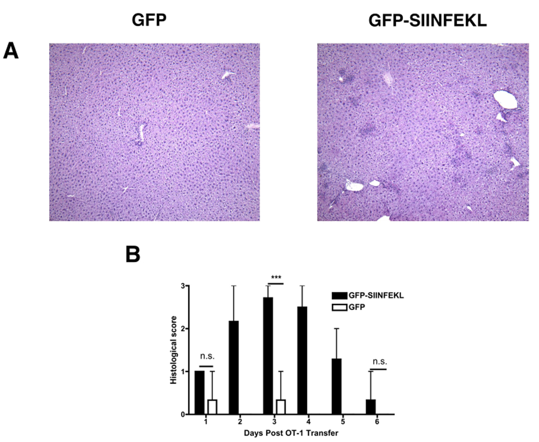
Figure 1. Histologic assessment of AAV-transduced livers.

C57BL6 mice were transduced with either rAAV2 GFP or rAAV2 GFP-SIINFEKL vectors by direct intrahepatic injection. All mice received 5×10⁶ OT-1 cells i.v. three weeks later. H&E-staining of the livers demonstrated the presence of numerous, discrete inflammatory foci in mice expressing the target antigen (A). The severity of liver injury was scored and plotted for 24-hr intervals post OT-1 transfer (B). Data points represent means ± range for n ≥ 3 animals per time point. ***, P < 0.0001; N.S, not significant. Data are the sum or representative of two independent experiments.
