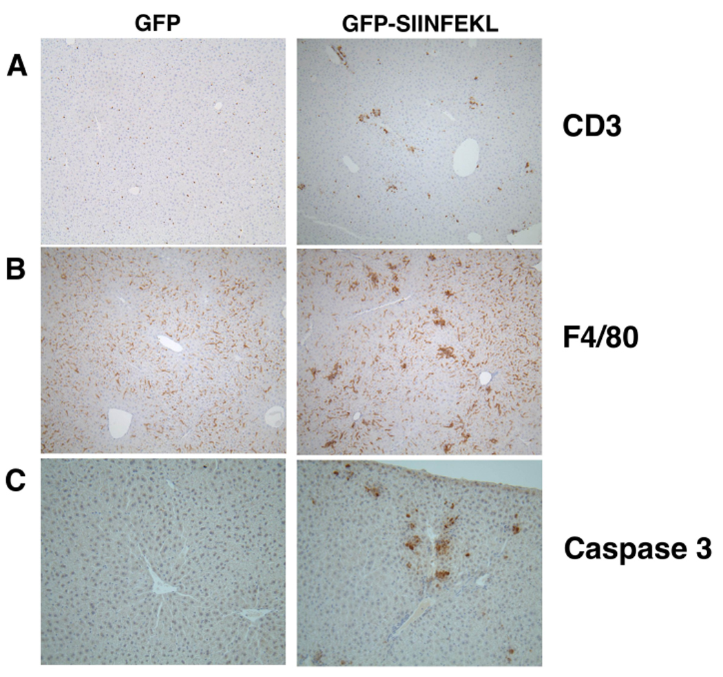
Figure 2. Histological staining for CD3, F4/80 and Caspase 3.

Liver sections from WT mice that had received rAAV2 GFP-SIINFEKL, or rAAV2 GFP, and OT-1 cells were stained for CD3 (A), F4/80 (B), or activated/cleaved Caspase 3 (C).

Liver sections from WT mice that had received rAAV2 GFP-SIINFEKL, or rAAV2 GFP, and OT-1 cells were stained for CD3 (A), F4/80 (B), or activated/cleaved Caspase 3 (C).