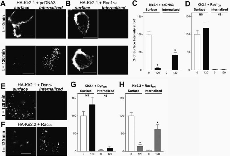
FIGURE 8. Rac1DN Inhibits Endocytosis of Kir2.1 Channels.
A-B) Representative confocal images from cells expressing an extracellularly HA-tagged Kir2.1 channel cDNA and either (A) pcDNA3 or (B) Rac1DN cDNA Cells were incubated with anti-HA antibodies on ice 15min and returned to 37° C for 0 (t=0), or 120 (t=120) minutes. Cells were then incubated with Alexa-647 conjugated antibodies prior to permeabilization to label channels on the plasma membrane (surface), and subsequently permeabilized and labed with Alexa-488 conjugated antibodies to visualize endocytosed channels (internalized). Rac1DN co-expressing cells failed to show internalization of Kir2.1 even after 120 minutes. D) Representative confocal images from cells expressing Kir2.2 with an HA tag in the extracellular region coexpressed with Rac1DN after 120 minutes of incubation. Note the intense labeling of internalized channels. Scale bars: 10μm. E-H) Bar graphs show quantification of surface and internalized channel intensity after 0, 60 or 120 minute incubation periods for cells expressing HA-Kir2.1 and either (E) pcDNA3, (F) Rac1DN or (G) DynDN, or (H) Kir2.2 and Rac1DN. Intensity is expressed as a percent of the total surface intensity at time 0. Experiments were repeated three separate times. N=5-14 cells per condition.

