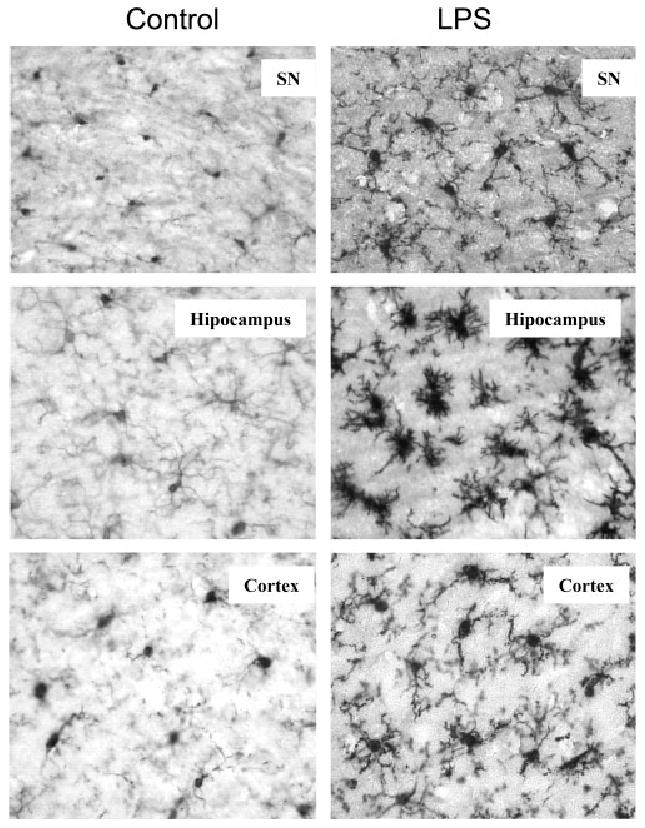
Fig. 3.

Immunocytochemical analysis of microglia. C57BL/6 mice were sacrificed 3 h following saline or LPS (5 mg/kg) i.p. injection. Brain sections were immnostained with Iba1 specific microglial antibody. Activated microglia in substantia nigra, hippocampus and cortex were shown by increased cell size, irregular shape, and intensified Iba1 staining in LPS-treated mouse brains. The images are from one experiment that is representative of three separate experiments.
