
Fig. 8.
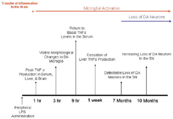
Systemic LPS causes chronic microglial activation and progressive dopaminergic neurotoxicity. This figure chronologically depicts the pro-inflammatory profiles in the periphery and the brain in response to LPS and the consequences for dopaminergic neuron survival. TNFα levels peaked in serum, liver, and the brain at 1 h, indicating that transfer of inflammation from the periphery to the brain was rapid. Changes in microglia morphology indicative of activation were present at 3 h. However, while peripheral inflammation (serum and liver TNFα production) had subsided by 9 h (serum) and 1 week (liver) after LPS treatment, brain TNFα and microglial activation remained elevated for up to 10 months. Interestingly, significant loss of dopaminergic neurons was first detected only at 7 months after treatment and increased in severity at 10 months after LPS exposure. These events document the development of inflammation in the substantia nigra in response to peripheral LPS administration and the consequent initiation of delayed and progressive dopaminergic neurotoxicity.