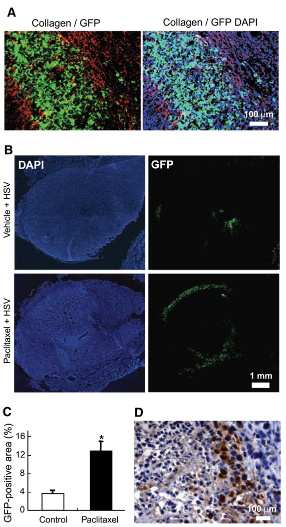
Figure 4.
Effect of paclitaxel pretreatment on viral distribution in collagen-rich MDA-MB-361HK tumors. A, collagen I immunostaining of frozen sections from tumors infected with HSV. The HSV infected area (GFP, green) was bordered by collagen fibers (Cy3, red). Note that there is less cell infection outside (DAPI staining, blue) the HSV infected area bordered by collagen fibers. B, HSV distribution in 361HK tumors. In all animals, the viral solution was injected in two different sites. In control tumors treated with vehicle, the HSV distribution was very limited and generally restricted to the tumor center. The HSV infection was located at the tumor edge in paclitaxel-treated tumors. C, quantification of viral distribution. Paclitaxel pretreatment significantly increased the viral distribution compared with control tumors (*, P = 0.003). D, HSV immunostaining of virus-infected areas. After paclitaxel pretreatment, HSV-infected cells were often located at the interface of necrotic and nonnecrotic tumor areas.

