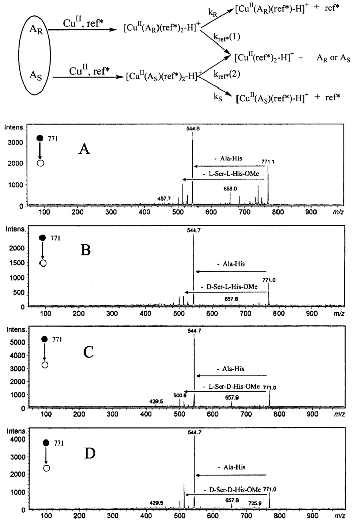
Fig. 7.

The top scheme illustrates the principle of kinetic method and the bottom multipart figure shows the CID fragmentation patterns of [CuII(A)(ref*)2-H]+ (m/z 771), with the ref* molecule being Ala-His dipeptide, and various analytes: (A) A = L-Ser-L-His-OMe; (B) D-Ser-L-His-OMe; (C) L-Ser-D-His-OMe ; and (D) D-Ser-D-His-OMe. Patterns of competitive collision for each stereoisomeric complex depend on their respective stabilities, therefore, the abundance ratio Re=[CuII(A)(ref*)-H]+ / [CuII(ref*)2-H]+ of different complexes can be used to measure the relative abundance of stereoisomers when a calibration curve is available. Top panel [119] and bottom panel [95] are adapted with permission from Wiley-Blackwell.
