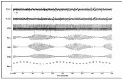
Figure 2.

Five minute section of a polysomnographic record of EEG (c3a1, c4a2), electrocardiogram (ECG), oronasal airflow (flow), abdominal ventilatory effort (ABD), thoracic ventilatory effort (THO), and oxygen saturation measured at the finger tip of the left hand (SaO2). Typical breathing pattern with Cheyne-Stokes respiration with hyperpnoeic and apnoeic sequences in sleep stage 2.
