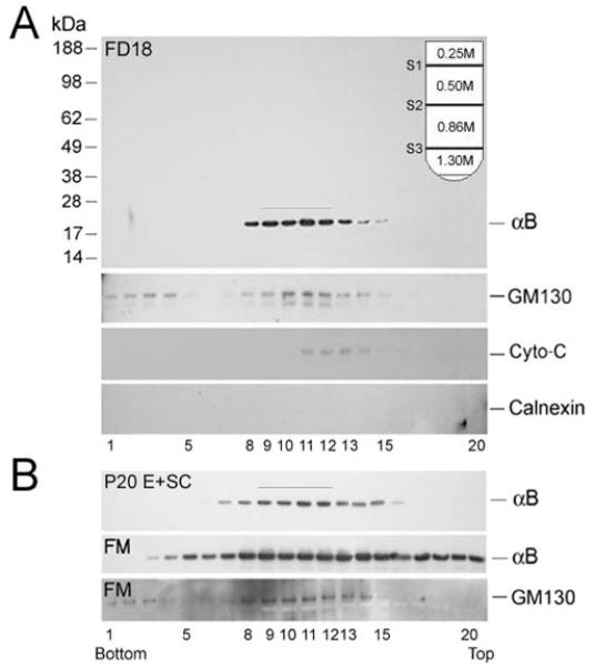
Figure 3.
αB-crystallin fractionates as a Golgi membrane- associated protein. (A) Distribution of αB-crystallin in FD18 whole lens postnuclear extracts (5.3 mg protein; top), fractionated on discontinuous density sucrose gradient (inset). (B) Distribution of αB-crystallin in postnuclear homogenates of E+SC (5.29 mg protein) and FM (6.53 mg protein) of P20 lens. For anti–αB-crystallin immunoblots, 1 μL was used from each fraction; for the rest, 10 μL were used. In the FD18 lens and the E+SC of the P20 lens, αB-crystallin predominantly fractionates with the Golgi membranes (fractions 9–12, bar). A whole immunoblot with molecular mass standards (kDa) is shown in FD18 panel; only relevant areas are shown in other panels.

