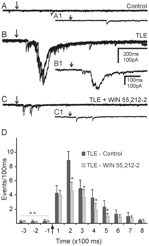
Figure 7. Cannabinoid-mediated attenuation of EPSC bursts evoked by glutamate ‘uncaging’ in the granule cell layer.
Sets of 3 overlapping traces showing evoked EPSCs after photolytic release of caged glutamate (250 µM) at the same location in the granule cell layer of control mice and mice with TLE. Arrows indicate the time of uncaging. A. Uncaging glutamate in the granule cell layer did not evoke a synaptic response in control mice. B. EPSC bursts evoked in pilocarpine-treated mice that survived SE. C. In the same cell as B, EPSC bursts were attenuated by WIN 55,212-2 (10 µM). A1, B1 and C1 show a single expanded trace from the group of traces shown in A, B and C. D. Grouped data from 6 cells showing the effect of WIN 55,212-2 on EPSC bursts evoked by photolysis of caged glutamate. Glutamate uncaging increased EPSC frequency over baseline; ** indicate significant difference in EPSC frequency before and after photolysis (p<0.05). * indicate a significant reduction in EPSC frequency after application of WIN 55,212-2 during comparable post-photolysis 100 ms time periods (p<0.05).

