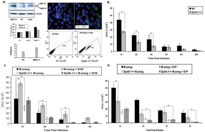
Figure 2. SphK-1 overexpression confers resistance to M. smegmatis infection in macrophages.
(A) Sphk-1 was overexpressed in macrophages and validated by western blot, immunofluorescence and competitive S1P titers in WT and SphK-1++ macrophages. (B) Sphk-1 overexpression confers resistance to infection. Both WT and SphK-1 ++macrophages were infected with M. smegmatis and mycobacterial killing was observed up to 24 h post infection. (C) The cells under section (B) were treated with DHS and the effect on M. smegmatis killing was again evaluated up to 24 h post infection. (D) S1P regulates mycobacterial growth in macrophages. The cells under section (B) were supplemented with S1P (5 µM) and the effect of S1P on mycobacterial infection was monitored during the first 4 h time period. Data are represented as mean of CFU ± SEM from three independent experiments. ** Indicates p≤0.01; * indicates p≤0.05.

