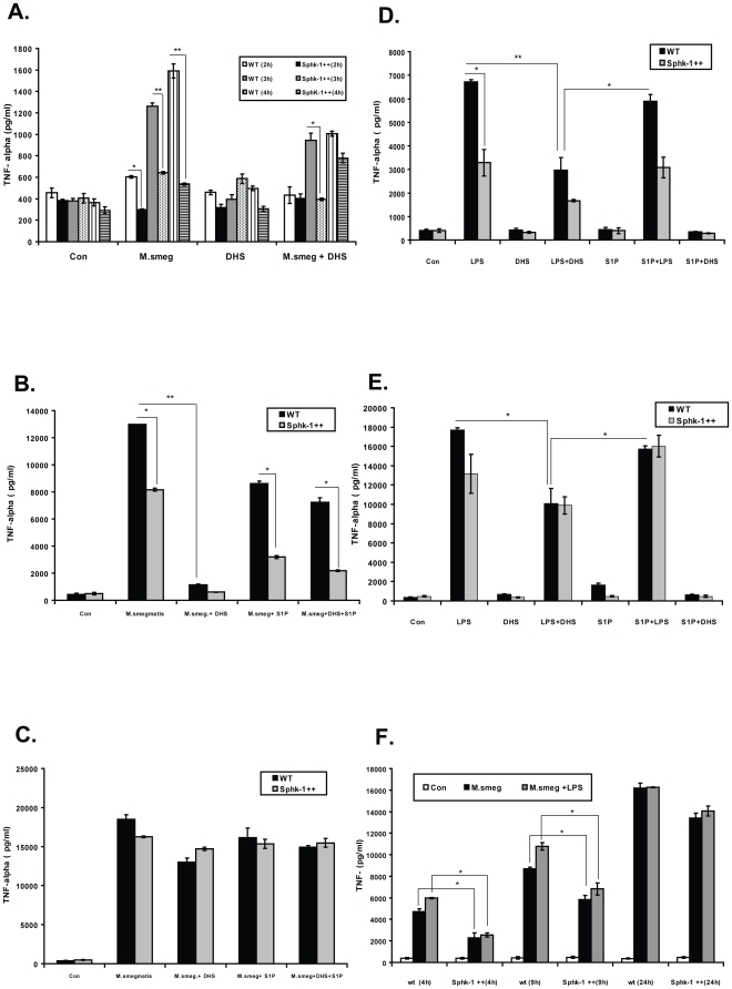
Figure 8. SphK-1 regulates TNF-α secretion in macrophages.
(A) Both WT and SphK-1++ macrophages were infected with M. smegmatis with and without DHS. The TNF-α titre was quantified in their culture supernatants until 4 h post infection. (B,C) Delay in the secretion of TNF-α by S1P in macrophages. The macrophages were infected with M. smegmatis with and without DHS (20 µM) and S1P (5 µM), and their culture supernatants were collected at 9 h (B) and 24 h (C) post infection and TNF-α titre was quantified. (D) The macrophages were stimulated by LPS with and without S1P and DHS, TNF titre was quantified at 9 h (D) and 24 h (E) post treatment. (F) Sphk-1 overexpression delays in the secretion of TNF-α by infected macrophage upon LPS co-stimulation. Both WT and Sphk-1++ macrophages were infected with M. smegmatis and co-stimulated with LPS for indicated time intervals. The TNF titre was quantified. Data are represented as a mean ρg/ml ± SEM from three independent experiments. ** Indicates p≤0.01; * indicates p≤0.05.

