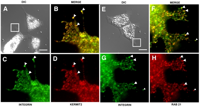
Figure 10. Colocalization of α5β1, kermit2, and Rab 21 in Xenopus embryonic cells.
Activin-treated embryonic cells were plated on FN substrates and stained for α5β1 (green), or kermit2 (red D), or Rab 21 (red H). (A, E) DIC images of adherent cells, boxes represent areas magnified in B–D and F–H. (A–D) Integrin (C; green) and kermit2 (D; red) colocalize at sites of adhesion (arrowheads in B–D). (E–H) Rab 21 (H; red) and integrin (G; green) colocalize in embryonic cells (arrowheads in F–H). Staining of cells with secondary antibodies alone produced no detectable signal. Size marker = 25 µM.

