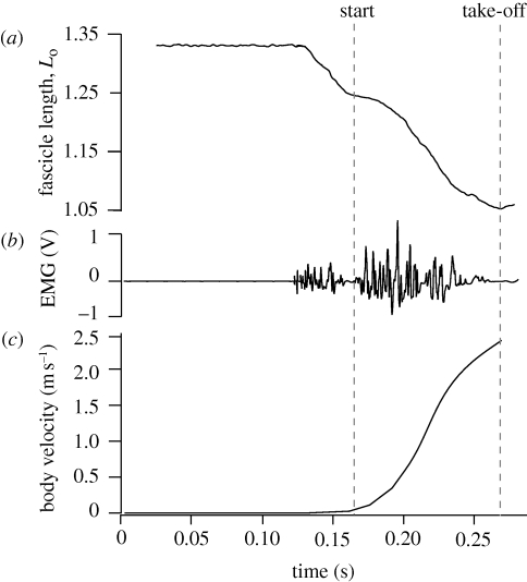
Figure 2.
Muscle length, EMG and body velocity for a representative jump. (a) Fascicle lengths measured by sonomicrometry normalized to the muscle's optimal length (Lo). (b) Muscle activity during jumps. The two separate bursts of EMG activity shown occurred in many but not all jumps. (c) Plot of the body velocity during the jump measured from high-speed video. This plot shows that little external movement occurred during the period of initial muscle activation and shortening. The beginning of the jump as measured from external video often coincided with a slowing of fascicle shortening and reduced EMG activity.

