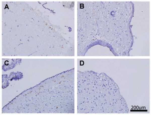
Fig. 3.

ADAMTS13 localizes to corpora amylacea (CA) in normal brain and multi-infarct dementia. Sections from normal (A, B) and vascular dementia (C, D) brains were stained for ADAMTS13, which showed a similar distribution of staining as thrombospondin1. Halo-shaped staining occurred in the cortex (A, D) and perivascular regions abutting the ependyma (B). Subependymal CA were strongly labeled as well (C).
