
Figure 5. Endogenous IRF3 and IRF7 RNA and protein expression is reduced during EBV lytic infection.
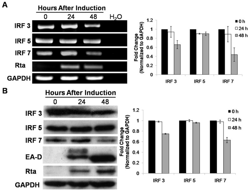
Viral reactivation was performed in Akata cells where the cells were incubated with F(AB′)2 fragment to human IgG for 0 hours (0 h), 24 hours (24 h), or 48 hours (48 h). A) Total RNA was harvested and semi-quantitative RT-PCR performed examining IRF3, IRF5, IRF7, and Rta RNA expression. B) Whole cell lysates were collected and Western blot analyses performed examining IRF3, IRF5, IRF7, Rta, and EA-D protein expression. Densitometry was performed in which relative RNA or protein expression was normalized to relative GAPDH expression. Data are shown as fold change (relative to 0 hour post induction) ± standard deviation of experiments performed in triplicate.