
Figure 6. Endogenous IFN-β RNA levels decrease during EBV lytic infection and coincide with increased susceptibility to Sendai virus infection.
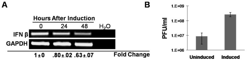
Viral reactivation was induced in Akata cells as in Figure 5. A) Total RNA was harvested and semi-quantitative RT-PCR performed to detect IFN-β and GAPDH RNA at 0, 24, and 48 hours post induction. Densitometry was performed where relative IFN-β levels were normalized to relative GAPDH levels. Data are shown as the fold change (relative to 0 hour post induction) ± standard deviation of experiments performed in triplicate. B) 24 hours after induction (or mock-induction), Akata cells were infected with Sendai virus (50 HA units/ml). 24 hours after infection, supernatant fluids were collected for plaque assays on LLC-MK2 cells. Viral titers were determined and data are shown as PFU/ml ± standard deviation of experiments performed in triplicate.