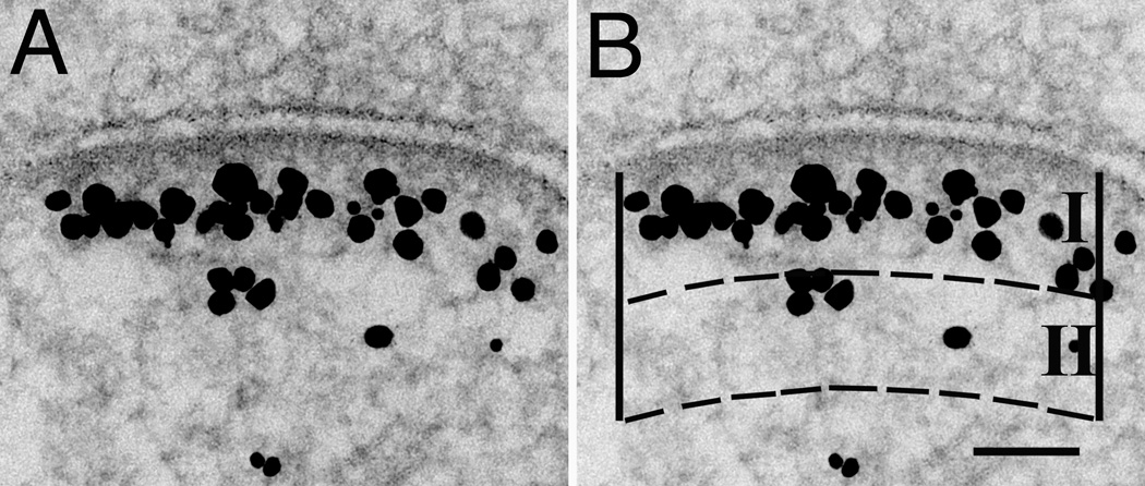
Figure 2.
Synapse labeled with pan Shank antibody (A) shown with superimposed mask (B) to illustrate the method for measuring the amount of label for Shanks in the vicinity of the PSD. Two zones, I and II, which are equal in area are delimited between arcs parallel to the PSD, and two vertical edges originating from the ends of the PSD. Zone I is designed to include the contiguous network immediately below the PSD. Each particle is assigned to the zone containing the majority of its areal projection. Scale bar = 100 nm.

