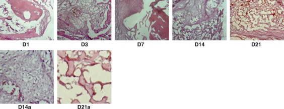
Fig. 1.
Histology of bone healing. Day 1: blood clot formed at the fracture end. Day 3: cell density increased in the clot as results of cell proliferation and local inflammation. Day 7: fibroblasts condensed at the fracture end. Day 14: callus formed at the fracture site with mixed tissues of fibrous, cartilaginous and bone. Day 14a: enlarged area of the cartilaginous tissue in the callus. Day 21: callus matured with mostly bone and bone marrow formed in the bone (Day 21a). Note: hematoxylin and eosin staining.

