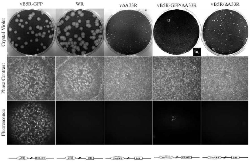
Fig. 1. Plaque phenotypes.

Confluent BS-C-1 cell monolayers were infected with the indicated viruses and overlaid with semi-solid media. Two days PI, phase contrast and fluorescence images were captured using a fluorescent microscope. Cell monolayers were stained with crystal violet three days PI and imaged. For vB5R-GFP/ΔA33R, insert shows an enlarged plaque in the boxed area. Schematic representation of the genome of each recombinant virus is shown below the fluorescence images.
