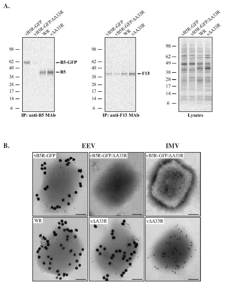
Fig. 6. A33 is required for efficient incorporation of B5-GFP into EEV.

RK13 cells were infected with the indicated viruses at a MOI of 10.0 and incubated with media containing [35S]-Met/Cys. 24 h PI, supernatants were collected, clarified, and loaded onto a 36% sucrose cushion to purify EEV. (A) Immunoprecipitation. Purified EEV were resuspended in RIPA buffer and equal amounts of radiation from each were subjected to immunoprecipitation with either an anti-B5 or anti-F13 MAb. Immune complexes were resolved by SDS-PAGE and radiolabeled proteins were detected by autoradiography. Equilibrated crude EEV lysates were analyzed to show that comparable amounts were used for each immunoprecipitation. The molecular weights in kDa and positions of marker proteins are shown. (B) Immunoelectron microscopy. Purified EEV and IMV were immunolabeled with an anti-B5 MAb and rabbit anti-L1 antibody, followed by 18 nm colloidal gold-conjugated goat anti-rat and 6 nm colloidal gold-conjugated goat anti-rabbit antibodies. Immunogold-labeled virions were negative-stained and visualized using a TEM. A representative virion from each is shown. Scale bars represent 50 nm.
