
Figure 4. NRG1 hypermethylation in the invasive and tumorigenic stages of the in vitro model and the estrogen receptor negative cell line MDA-MB-468.

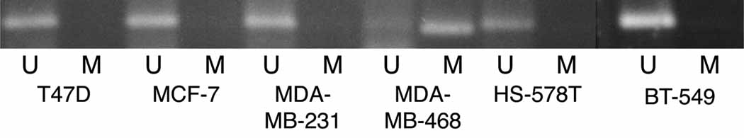
NRG1 methylation was studied by methylation specific PCR (MSP) in: A) the in vitro model of breast cancer progression and, B) in the human breast cancer cell lines T47D and MCF-7, both ERα positive and, the ERα negative cells MDA-MB-231, MDA-MB-468, HS578T and BT549. A band corresponding to NRG1 exon 1 was identified with two sets of primers, one pair recognized a sequence in which CpG sites present in the primer region were unmethylated (bisulfite modified to UpG), and the other recognized a sequence in which CpG sites were methylated (unmodified by bisulfite treatment). A representative experiment from three independent ones is showed.