Figure 1.
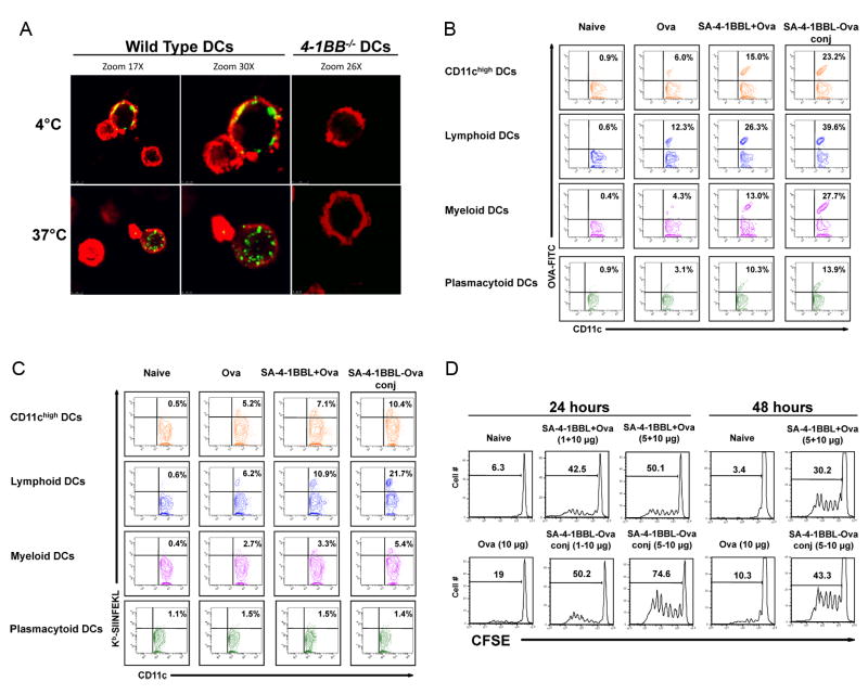
SA-4-1BBL targets conjugated antigen in vivo to DCs for enhanced uptake and cross-presentation. A, Bone marrow-derived DCs internalize SA-4-1BBL following 4-1BB binding. DCs from wild type and 4-1BB-/- C57BL/6 mice were incubated with SA-4-1BBL-FITC at 4°C for receptor binding, washed, and then incubated for 1 h at 4°C or 37°C for internalization. CD11c (red) DCs from wild type, but not 4-1BB-/-, mice internalize SA-4-1BBL (green) at 37°C, but not 4°C. A minimum of 3 fields per slide were analyzed. B, SA-4-1BBL-Ova conjugates enhance antigen uptake by DCs in vivo. C57BL/6 mice were injected s.c. with conjugated SA-4-1BBL-Ova-FITC (25-10 μg), nonconjugated SA-4-1BBL+Ova-FITC (25+10 μg), Ova-FITC alone (10 μg), or left untreated. Draining lymph node cells were harvested 24 h later, stained with CD11c-PE, CD11b-PErcp-Cy5.5, B220-PEcy7, and CD8α-APC-cy7 Abs, and analyzed in flow cytometry. Data for Panels A and B are representative of two independent experiments. C, SA-4-1BBL-Ova conjugates increase antigen cross-presentation by DCs in vivo. C57BL/6 mice were vaccinated as in B, except Ova without FITC was used as antigen. Draining lymph node cells were harvested 24 h later and subjected to Ab staining as in B. APC-conjugated 25D1.16 Ab was used to detect Kb/SIINFEKL on the surface of DCs. D, SA-4-1BBL-Ova conjugates result in increased antigen presentation to OT-I CD8+ T cells in vivo. C57BL/6.SJL (CD45.1+) mice were immunized s.c. with Ova (10 μg) conjugated or nonconjugated with various doses (μg) of SA-4-1BBL as indicated. 2×10⁶ sorted OT-I T cells (CD45.2+) were labeled with CFSE and injected i.v. 24 or 48 h later. Proliferation was assessed using flow cytometry 3 days later. Data are representative of a minimum of three independent experiments for Panels C and D.

