Figure 1.
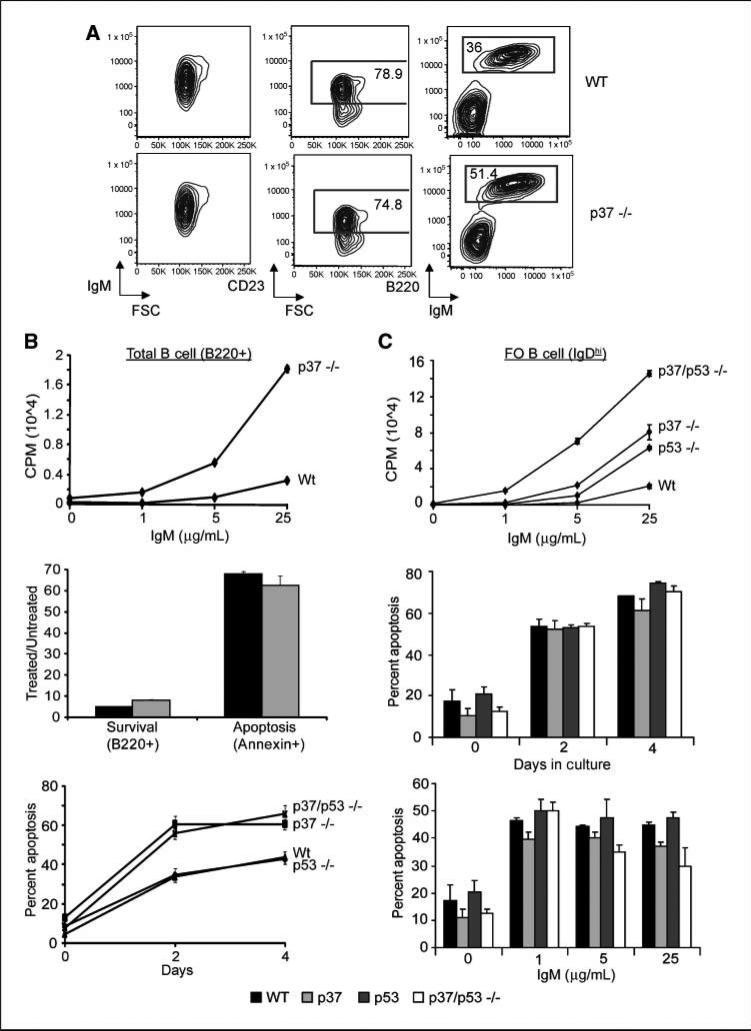
Proliferation and apoptosis in p37-null and p37/p53-double null total B cells and FO B cells. A, FACS analysis of spleen cells from a healthy 8-mo-old wt control mouse and matched p37-null mouse. FSC is forward scatter (proportional to cell size). Left and right plots, all live spleen cells within the lymph scatter gate; middle plots, data from CD23+ B cells. B, [3H] thymidine uptake assay of Wt, p53-null, p37-null, and p37/p53-double null purified total B cells (CD45R/B220+; top). Assay was performed in triplicate and repeated twice. B cells were treated with the indicated amount of anti-IgM and harvested 24 h for [3H] thymidine uptake assay. Middle, mice were treated with 10Gy ionizing radiation and spleens were harvested 8 h later. Single cell suspensions were then stained with B220, Annexin V, and 7-AAD before being analyzed by FACS. Shown is the ratio of treated versus untreated cells that were positive for B220 or Annexin V. Experiment was done in triplicate and repeated at least thrice. Bottom, apoptosis assay on purified total B cells (B220+) from Wt, p53-null, p37-null, and p37/p53-double null mice treated with 100 nmol/L dexamethasone. Plots show percent of sub-G1. Assay was done in triplicate and repeated twice. C, [3H] thymidine uptake assay of Wt, p53-null, p37-null, and p37/p53-double null–purified FO B cells (IgDhi; top). Assay was done as in B. Middle and bottom, apoptosis assay on purified FO B cells (IgDhi) from wt, p53-null, p37-null, and p37/p53-double null mice. FO B cells were either left in prolonged culture for 4 d, with cells being harvested and stained with propidium iodide every other day, or FO B cells were treated with increasing amounts of IgM and harvested 16 h later. Plots show percent of sub-G1. Assays were done in triplicate and repeated twice.

