Figure 3.
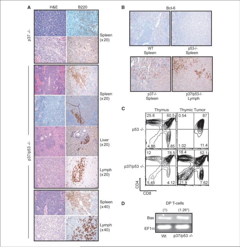
Characterization of tumors from p37/p53-double null mice. A, representative H&E or CD45R/B220-stained tumor sections from p37-null or p37/p53-double null mice. The p37/p53-double null liver, lymph node, and first spleen sections are from one mouse. All pictures were taken at ×20 magnification unless otherwise indicated. B, representative Bcl-6 stained wt, p53-null, p37-null, and p37/p53-double null spleenic or lymph node tumors. All pictures were taken at ×20 magnification. C, FACS plot of CD4 and CD8-stained thymic tumors from p53-null or p37/p53-double null mice. D, relative fold induction of Bax expression by quantitative real-time RT-PCR on CD4/CD8 DP sorted T cells from wt or p37/p53-double null thymii.

