
Figure 1.
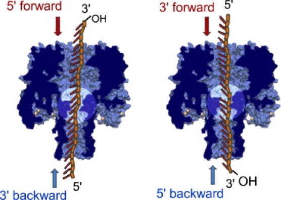
Schematic representations of the DNA inside the pore for both DNA orientations and both translocation directions. The tilt of the bases is purely schematic to illustrate the MD simulation results obtained in Mathé et al. (11). Note the equivalence of the static situations for DNA with their 5′ end first in the forward direction (respectively, 3′ end first in the forward direction) and DNA with their 3′ end first in the backward direction (respectively, 5′ end first in the backward direction).