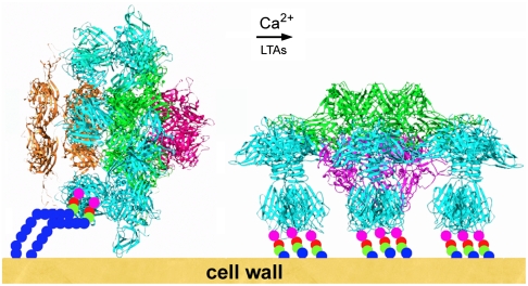
Fig. 4.
Schematic representation of the putative adsorption mechanism of phage p2 to its host. The rest form (left) of the baseplate is activated by Ca2+ cations and by the traction of lipoteichoic acids bound to a few receptor-binding sites which destabilize the RBPs. They subsequently rotate by 200° and become available for a complete binding. Concomitantly, the tip of the baseplate opens, giving way to dsDNA.

