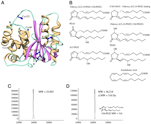
Fig. 1.
3D structure of UCH-L1, chemical structures of prostaglandins and ESI-MS analysis of UCH-L1 modification by 15d-PGJ2. (A) Crystal structure of human UCH-L1 (PDB:2ETL) (9) in ribbon representation (strands: Magenta; helices: Yellow; irregular loops: Teal). The a-helical and b-sheet domains are located on the left and right hand side of the active site cleft, respectively. Side chains for the six Cys residues are depicted in purple ball and stick representation. (B) Prostaglandins used in the present study; molecules containing the α,β-unsaturated carbonyl moiety in the cyclopentenone ring are depicted on the left, those with saturated rings on the right, and arachidonic acid at the bottom. (C–D) ESI-MS analysis of free (C) and 15d-PGJ2 conjugated UCH-L1 (D).

