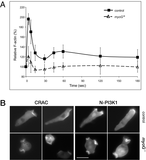
Fig. 4.
Chemotactic signaling is impaired in myoG null cells. (A) Actin polymerization in response to cAMP is dampened in myoG- cells. Analysis of changes in cytoskeletal F-actin levels in both control (■) and myoG mutant (△) cells after a 100-nM cAMP pulse (administered at time = 0). Data shown from three independent experiments represent the mean ± SD of control (n = 8) and myoG null cells (n = 14). (B) Localization of CRAC-GFP and N-PI3K1-GFP in a cAMP gradient. Aggregation competent cells were exposed to a cAMP gradient in a Zigmond chamber and photographed after ~15 min. The cAMP source is at the right in all panels. (Scale bar, 10 μm.)

