Fig. 3.
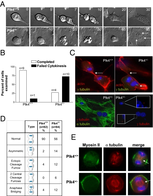
Plk4 is required for myosin II localization and completion of cytokinesis. (A) Time-lapse DIC microscopy of mitotic P3 MEFs, showing failure of cytokinesis in a bipolar Plk4+/− cell, resulting in multinucleation (arrows indicate nuclei, time points are in minutes). (B) Proportion of bipolar mitotic P3 Plk4+/+ and Plk4+/− MEFs completing and failing cytokinesis. (C) (Upper) P3 Plk4+/+ and Plk4+/− MEFs in late mitosis stained for F-actin (red), γ-tubulin (green), and DNA (blue). White arrows indicate cleavage furrow formation. (Lower) P3 Plk4+/− MEF in late mitosis stained for DNA (blue) and α-tubulin (green) and/or γ-tubulin (red). (Insets) A single γ-tubulin–positive centrosome is seen at each pole. (D) Summary of mitotic phenotypes in P3 Plk4 MEFs. Plk4+/− MEFs frequently undergo asymmetric division and ectopic cleavage furrow formation. (E) Representative immunofluorescent images of MEFs in cytokinesis showing mislocalized myosin II in Plk4+/− cells (arrows indicate midbody location).

