Fig. 3.
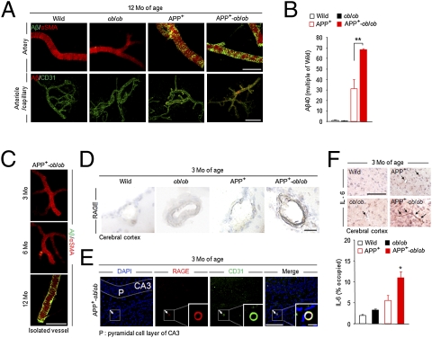
Increased vascular amyloid deposition and inflammation in APP+-ob/ob mouse brain. (A) Immunohistochemical detection of Aβ40 deposition in isolated brain microvessels of 12-month-old mice. Brain microvessels were also stained for anti–α-smooth muscle actin (a vascular smooth muscle cell marker) and CD31 (endothelial cell marker). (Scale bar, 100 μm.) (B) Quantification of Aβ40 level in isolated brain microvessels by ELISA (n = 3 per group). **P < 0.01. (C) Cerebrovascular amyloid deposition in APP+-ob/ob mice was age-dependent and appeared after 6 months of age. (Scale bar, 100 μm.) (D) Immunohistochemical staining for RAGE in brain sections of young (3-month-old) mice. Strong immunoreactivity was detected in brain vessels (cerebral cortex) of APP+-ob/ob mice. (Scale bar, 30 μm.) (E) Brain section of 3-month-old APP+-ob/ob mouse immunolabeled for RAGE and CD31 and counterstained with DAPI. Colocalization of RAGE and CD31 in cerebral vessel is denoted by arrow and magnified (Inset). (Scale bars, 100 μm; 10 μm for Inset.) (F) IL-6–positive microvessels (cerebral cortex) in 3-month-old mouse (Left) and quantitative image analysis of IL-6–positive vessels (percent occupied; Right, n = 3). (Scale bar, 100 μm.) *P < 0.05 for APP+-ob/ob mice versus other genotypes.

