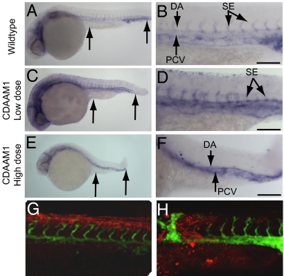
Fig. 4.
CDAAM1 inhibits angiogenesis in zebrafish. (A and B) WT embryos demonstrating normal Fli-1 expression. (C and D) Embryos injected with 2 ng of CDAAM1 RNA. (E and F) Embryos injected with 4 ng of CDAAM1 RNA. A, C, and E (arrows) reveal the A-P length as readout for the PCP pathway. B, D, and F display vascular endothelial cell staining in the somite region. (G and H) Confocal images with Fli1-EGFP expression overlaid on lineage tracer identifying transplant location. Zebrafish embryo manipulation, RNA preparation, and imaging are described in Materials and Methods. (Scale bars = 100 μm.)

