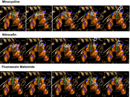
Fig. 3.
Top five binding modes of minocycline (Top), nitrocefin (Middle), and fluorescein maleimide (Bottom) for the Binding protomer of AcrB (2DRD) predicted by Autodock Vina. The view in this figure, as well as in Fig. 4 and Figs. S1 and S2, is the side view of the binding pocket (shown as a surface with carbons in orange), similar to Fig. 2B Inset. It is from the outside of the Access protomer, which was removed by clipping together with much of the Extrusion and Binding protomers. The computer-predicted poses of the ligands are shown in stick models in CPK colors, and the minocycline in the crystal structure (2DRD) is shown for reference in a stick model with carbons in green. The poses are arranged in the order of calculated binding energy, beginning with the one predicted to be most stable at extreme left.

