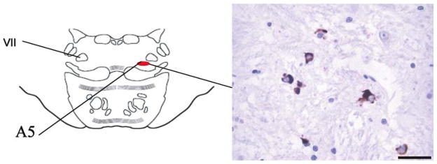
Fig. 3.
Six micrometer paraffin-embedded sections of the pons at the level of the A5 area (left) stained for α-synuclein and showing the distribution of glial cytoplasmic inclusions in a 68-year-old man with pathological diagnosis of multiple system atrophy (right) (MSA, postmortem delay 23 h). Bar 25 μm

