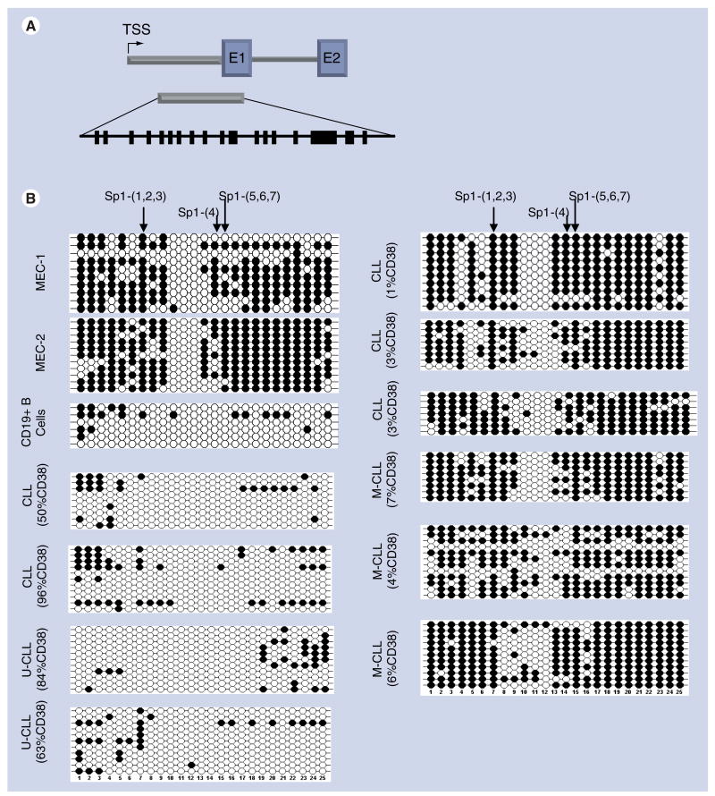
Figure 3. Bisulfite sequencing of DLEU7.
DNA methylation of 25 CG dinucleotides was examined in a 5′ region of DLEU7 that spans part of a region spanning the predicted 5′UTR and part of the first exon; the location of this region with respect to the TSS is shown. The CG dinucleotides are shown as heavy black lines. The methylation status of 25 CG dinucleotides was determined from bisulfite-treated DNA of CLL patients with different CD38 expression levels and either M-CLL or unmutated U-CLL IgVH mutational status, two CLL cell lines (MEC1 and MEC2) and CD19+ B cells from a healthy donor. Each row is the result from an individual clone across the 25 CG dinucleotides. Filled circles indicate methylated cytosine and open circles are unmethylated sites.
CLL: Chronic lymphocytic leukemia; M-CLL: Mutated CLL; TSS: Transcription start site; U-CLL: Unmutated CLL.

