
Fig. 5.
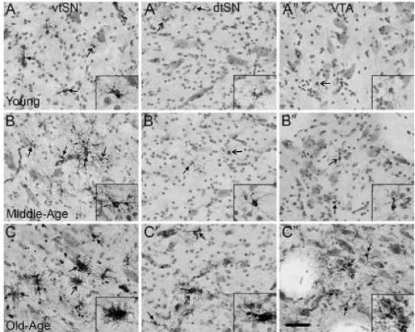
Normal aging is associated with a chronic state of moderate inflammation in the midbrain, and the vtSN is most severely affected. (A-A”) In young animals, the vtSN (A), dtSN (A’), and VTA (A”) have microglia exhibiting resting morphologies (images taken from an animal with − / − / − rating). (B-B”) In middle-age animals, the vtSN contains microglia undergoing early stages of activation as evidenced by increased HLA-DR staining and hyper-ramified morphologies (B), while microglia in the dtSN (B’) and VTA (B”) continue to exhibit resting morphologies (images taken from an animal with + / − / − rating). (C-C”) In the midbrain of aged animals, there is a ubiquitous presence of hyper-ramified microglia in the vtSN (C), dtSN (C’), and VTA (C”; images taken from an animal with ++ / + / + rating). (C) Only within the vtSN of an old-age animal (and one middle-age animal – not shown) were the majority of microglia found to exhibit morphological characteristics consistent with advanced stages of activation. Close arrows indicate HLA-DR+ microglia and open arrows indicate cells in insets. Scale bar: (A-C”) 50 μm.