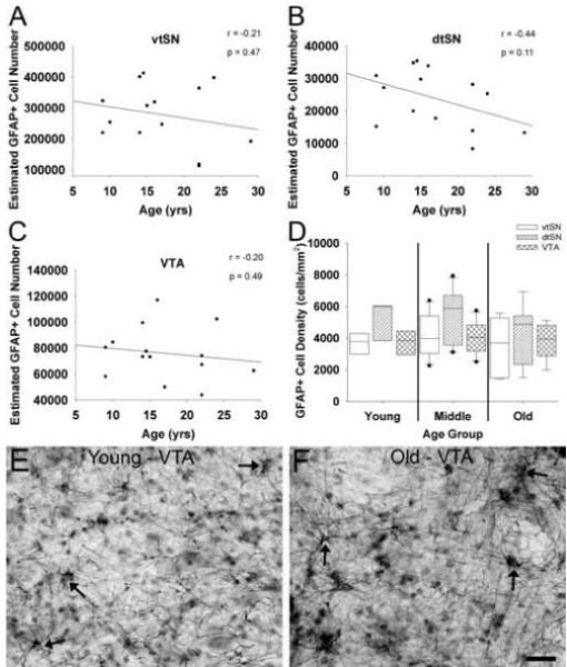
Fig. 6.

Aging is not associated with increased numbers of GFAP+ astrocyte in DA midbrain subregions. (A-C) Increasing chronological age was not significantly correlated with the number of GFAP+ astrocytes in the vtSN (A), dtSN (B), or VTA (C). (D) Astrocyte density was similar in the vtSN, dtSN, and VTA of all age groups. (E and F) The photomicrographs depict no change in astrocyte density in the VTA of a young (E) and old-age (F) animal. Arrows indicate GFAP+ astrocytes. Scale bar: (E and F) 25 μm.
