
Figure 1. Flow cytometry analysis to demonstrate the expression of murine MHC molecules in CIITA-transfected DC-1 cells.
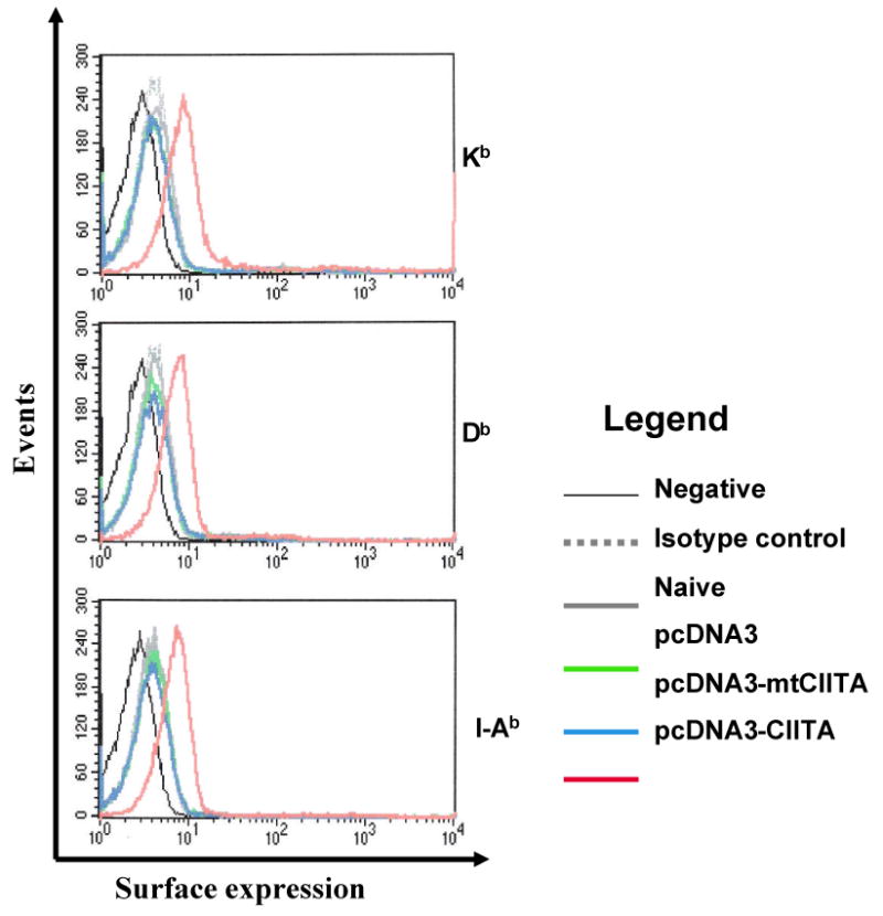
Flow cytometry data demonstrating the levels of H-2 Kb, H-2 Db, and I-Ab expression in CIITA-transfected cells and controls. The legend shows the vector plasmids with which the DCs were transfected. An immortalized dendritic cell line (DC-1) was transfected with CIITA or mutant CIITA (mtCIITA). Untransfected cells and cells transfected with the pcDNA3 vector backbone were used as a control. The expression of MHC I and II molecules was characterized using antibodies to MHC I H-2 Kb, H-2 Db, and MHC II I-Ab by flow cytometry analysis.