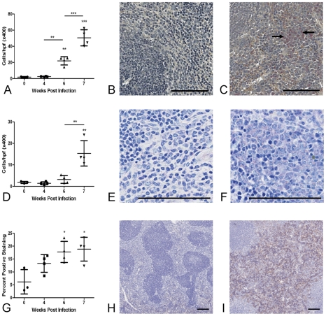
Figure 2. Splenomegaly is associated with accumulation of neutrophils, eosinophils and F4/80+ macrophages in the red pulp.
There was a significant increases in the number of neutrophils (A–C: Leder stain for neutrophils (pink stain, arrowed) ×200 in uninfected control mice (B) and at 7 weeks p.i. (C)), eosinophils (D–F, Giemsa stain ×400 in uninfected control mice (E) and at 7 weeks p.i. (F)) and F4/80+ macrophages (G–I: F4/80 staining in uninfected control mice (H) and at 7 weeks p.i (I). (red-brown) ×40) in the splenic red pulp from as early as 6 weeks p.i. Values represent mean cells/high power field (eosinophils and neutrophils) or percent positive staining (F4/80) ±1SD of mice pooled for microarray analysis (n = 4 per group). Bar = 100µm.

