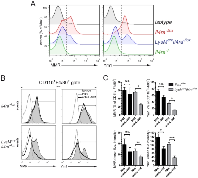
Figure 5. IL-10 signalling drives mannose receptor and Ym1 expression in macrophages independently of their IL-4Rα expression.
Peritoneal cells were harvested 7 days after injection of 3000 S. mansoni purified eggs in the peritoneum of Il4ra−/lox, LysMcreIl4ra−/loxor Il4ra−/− mice. (A) Representative histograms of macrophage mannose receptor (MMR) or Ym1 expression by peritoneal macrophages gated on FSChighSSClowCD11b+F4/80+ cells after exclusion of peritoneal eosinophils (FSClowSSChighCD11b+F4/80+) [37]. Black, isotype control; red, Il4ra−/lox; blue, LysMcreIl4ra−/lox; green, Il4ra−/−. Broken lines show threshold for positive signal. Data are representative of two independent experiments of pooled samples (n = 3). (B) Anti-IL-10 receptor treatment. Four mice out of eight in each group received 4µg of anti-IL-10R i.p. at day 0, 4 and 6 post-injection. Overlays of representative monoparametric histograms of macrophage mannose receptor (MMR) or Ym1 expression by peritoneal macrophages are shown as described in A. Bracketed line indicates positive signal. Dotted line, isotype control; greyscale, untreated mice; bold line, mice treated with anti-IL-10R. (C) Percent and mean fluorescent intensities of peritoneal macrophages expressing MMR or Ym1 based on flow cytometry analysis in B. Data is representative of two independent experiments with mean±SEM (n = 3). *p<0.05, ** p<0.01, *** p<0.001.

