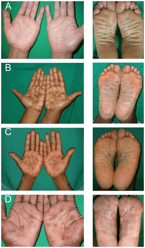
Figure 2. Palmar and plantar rash of secondary syphilis.
Typical palmar and plantar rash of secondary syphilis is shown in the representative figures. Similar lesions were evident in 59.6% of all secondary syphilis subjects enrolled. These lesions consist of smooth or scaly plaques and papules, which can become hyperpigmented in dark-skinned individuals as shown in the figure.

