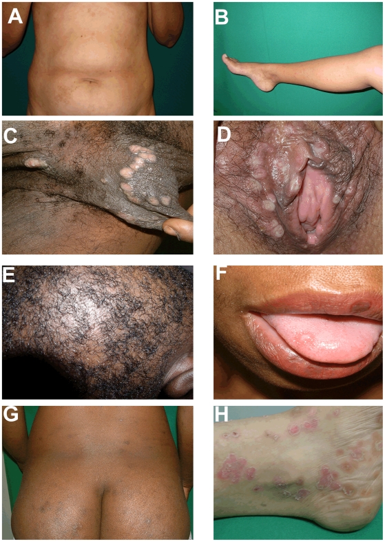
Figure 3. Mucosal and cutaneous lesions of secondary syphilis.
Secondary syphilis has been known as the “Great Imitator” due to the diversity of dermatologic lesions and which can be confounded with other cutaneous diseases. (A and B) Diffuse erythematous papular exanthem is shown over abdomen in A and lower extremity in B. (C and D) Multiple moist, hypopigmented, flattened plaques consistent with condyloma lata are shown on external genital areas (male and female respectively in C and D). (E) Inflammatory responses can affect hair follicles leading to “moth-eaten alopecia” as depicted in the figure. (F) Oral mucosal patches, as shown in the figure can be present during secondary syphilis. (G) Pigmentary plaques are shown over the buttocks of a dark-skinned secondary syphilis patient. (H) Psoriasiform syphilitic lesions, as shown in the representative micrograph, could easily be misdiagnosed as psoriasis.

