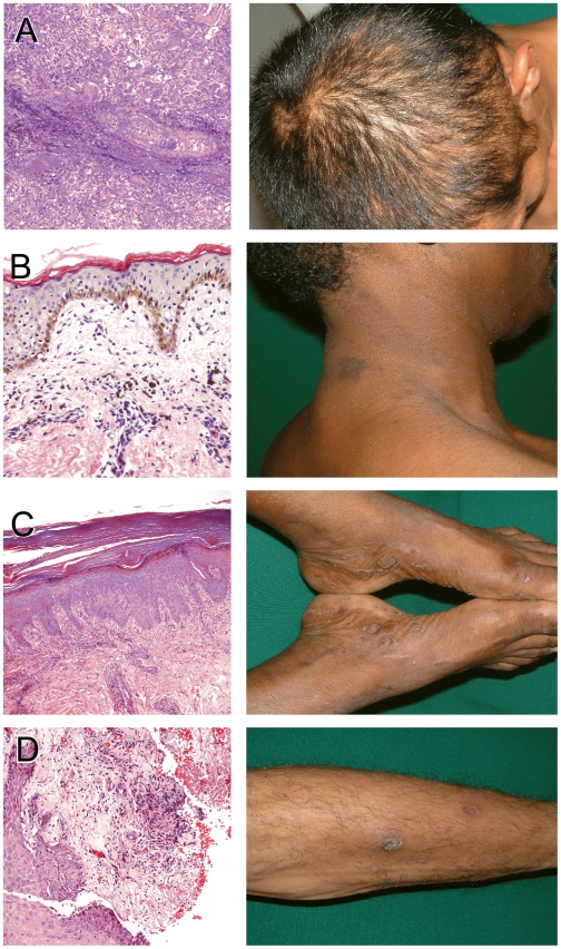
Figure 4. Secondary syphilis histopathology.
The figure shows histopathologic anomalies seen in punch biopsies obtained from four secondary syphilis patients skin lesions. Corresponding clinical appearance of the lesions are also shown. (A) Markedly inflamed hair follicle (“folliculitis”) with extension of inflammatory cell infiltrate into parafollicular blood vessel and connective tissue. Corresponding “moth-eaten” alopecia is shown in the adjacent micrograph. (B) Dark-skinned patient (pigmented basal keratinocytes); further darkening of a patch of skin in the form of a macule, as shown herein, is caused by deposition of dermal melanophages (“pigment incontinence”) (C) Skin biopsy obtained near the sole reveals a thick stratum corneum layer, epidermal reactive psoriasiform hyperplasia associated with chronic inflammation of the dermal papilla, and a superficial perivascular lymphoplasmocytic infiltrate. (D) Edge of an ulcer located in the lower extremity reveals fibrinoid exudate on the ulcer bed, surrounded by granulation tissue and reactive hyperplasia of the epidermis.

