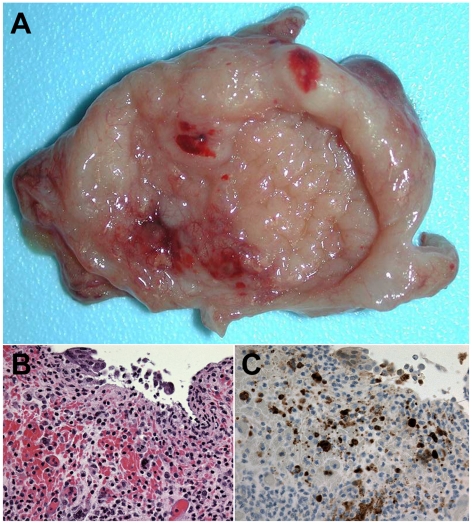
Figure 3. NiV infection and pathogenesis in the urinary bladder.
Subject 3 that succumbed on day 11 after i.t. and oral exposure to 8.1×10⁴ pfu of NiV. (A) Petechial to ecchymotic hemorrhages on mucosal surface of urinary bladder of animal of animal at necropsy. (B) Bladder ulcer showing inflammation and hemorrhage by H&E staining; 400× magnification. (C) Bladder ulcer by immunohistochemistry staining of NiV antigen; 400× magnification.

