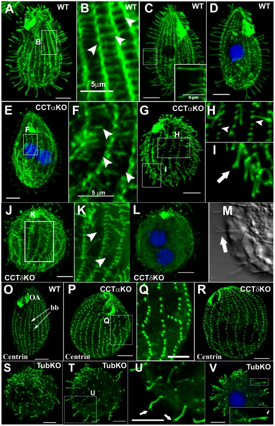
Figure 1. CCT subunits are required for assembly of axonemal, cortical and cell body microtubules.
Confocal immunofluorescence of α-tubulin (A to L and S to V) and centrin (O to R) in wildtype, CCTα-KO, CCTδ-KO and tubulin-KO Tetrahymena. In some images, DNA is stained with TO-PRO-3. (A–D) Wildtype cells. A higher magnification of the cortical region boxed in A is shown in B; In panel C, the inset shows a higher magnification of a group of cilia of the boxed area. (E–I) CCTα-KO cells 26 hpm. F represents a higher magnification of a boxed region of the cell cortex shown in E. H and I are higher magnifications of boxed regions shown in G. Arrowheads in F and H show either shortening or absent TMs. (J–M) CCTδ-KO cells 26 hpm. K shows a higher magnification of an area boxed in J. Arrowheads in K show shortening TM bundles. (M) A differential interference image of a portion of CCTδ-KO cell. The arrow points at a branched ciliary tip. (O–Q) Anti-centrin staining of respectively WT, CCTα-KO and CCTδ-KO showing disorganization of ciliary rows in CCT depleted cells; (Q) shows a higher magnification of a boxed region from cell shown in (P), depleted from CCTα-KO, where it is observed a variation in the distance between two consecutive basal bodies and presence of gaps reflecting absence of basal bodies in the row. (S–V) Tubulin-KO cells 26 hpm stained with antibody directed to α-tubulin. In (U), higher magnification of area boxed in (T), and inset in V arrows point at branched ciliary tips. Scale bar represents 10 µm except if mentioned differently.

