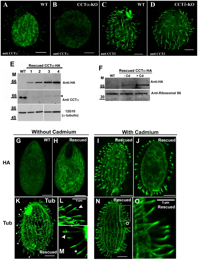
Figure 2. CCTαp-HA rescues CCTα-KO, localizes in cilia and its abundance affects cilia tips.
A–D) Confocal immunofluorescence with antibody directed to CCTα (A and B) and CCTδ (C and D) in wildtype, CCTα-KO and CCTδ-KO Tetrahymena cells showing that depletion of CCT genes reduces CCT levels in cell body and cilia. All images were taken with exactly under the same exposure conditions. (E) Western blots of total proteins obtained from wildtype (WT) and rescued CCTα-KO cells expressing CCTαp-HA under cadmium-inducible promoter probed either with anti-HA, anti-CCTα (affinity-purified) antibodies, or the anti-α-tubulin 12G10 antibody. Lane 1 and 3- cells grown in SPPA with 1.5 µg/ml of cadmium chloride for 24 and 48 h, respectively; lane 2 and 4- cells grown in SPPA with 2.5 µg/ml of cadmium chloride for 24 and 48 h, respectively. (F) Western blots of total proteins of WT and rescued CCTαp-HA cells, grown in SPPCT medium in absence (−) or presence (+) of cadmium (Cd) probed with anti-HA and anti-ribosomal S6 antibodies. (G–O) Epitope-tagged CCTαp localizes to cilia and its levels affect the integrity of ciliary tips. Tetrahymena CCTα-KO cells were rescued by introduction of a transgene expressing CCTαp-HA under MTT1 promoter, grown in the absence or presence of cadmium, and processed for immunofluorescence using anti-HA (G to J) and anti-α-tubulin (12G10) (K to O) antibodies. (G) A wildtype cell stained with anti-HA antibody. (H–J) A CCTαp-HA-expressing rescued cells stained with anti-HA antibodies and grown in SPPCT medium for 76 h, without cadmium (H) and with 2.5 µg/ml of cadmium (I and J); J shows an internal optical section of the cell shown in I. (K–O) CCTαp-HA rescued cells stained by anti-α-tubulin antibody that were grown in SPPCT medium for 76 hr either without cadmium (K–M), or with 2.5 µg/ml of cadmium (N–O). (L and M) Higher magnification of ciliary regions of cells grown in SPPCT without cadmium to show the abnormal cilia tip. (O) A higher magnification of a boxed region from cell shown in (N). Scale bar = 10 µm, except if mentioned differently.

