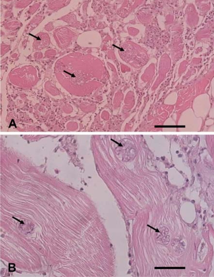
Fig. 1.
Optical micrographs of the muscle tissue infected with Heterosporis anguillarum. The tissue was cross-sectioned (A) and sectioned longitudinally (B). Multifocal sporocysts and hyaline degeneration are shown in the muscle fibers (arrowheads) and diffused severe lymphocytic inflammation can be seen in the muscle tissue. bar = 60 µm (A), 30 µm (B).

