Figure 1.
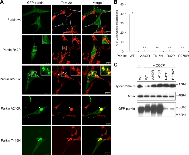
Disease-associated Parkin mutations are defective in mitophagy. (A) MEFs were transfected with GFP-tagged wild-type (WT) or mutant Parkin expression plasmid followed by an 18-h treatment of CCCP. Cells are immunostained with a Tom20 antibody to visualize mitochondria (red). GFP-parkin–transfected cells are marked by dotted lines. Arrows indicate parkin-positive mitochondria or mitochondrial aggregates. Bar, 10 µm. (B) The average percentages of mitochondria-free cells from three independent experiments from A are presented with standard deviation as error bar. **, P < 0.01 (C) MEFs were transfected and treated with CCCP as described in A, followed by an immunoblotting analysis with antibodies for cytochrome c, actin, and parkin. Note that levels of parkin R275W and R42P mutant were lower, as previously reported (Wang et al., 2005).

