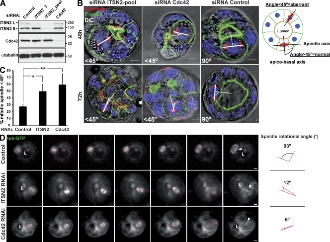
Figure 4.
Cells silenced for Cdc42 or ITSN2 present spindle orientation defects. (A) Down-regulation of ITSN2 and Cdc42 by siRNA. Cells were transfected with Cdc42, ITSN2, or control siRNA and allowed to form cysts for 72 h, and then total cell lysates were Western blotted for Cdc42 and α-tubulin (control). Molecular mass is indicated in kilodaltons. (B) Effect of Cdc42 and ITSN2 siRNA in spindle orientation. Cells were transfected with ITSN2 siRNA pool (left), Cdc42 siRNA (middle), or control siRNA (right) and plated to form cysts for 48 (top) or 72 h (bottom). Cells were stained to detect actin, acetylated tubulin (Ace tub), and chromatin (blue). The apicobasal axis (thin line) and the spindle axis (thick line) are drawn in white. (C) Quantification of misoriented spindles in 48-h cysts silenced with Cdc42, ITSN2, or control siRNA. The angle between the apicobasal axis and spindle axis was measured. Angles <45° were counted as abnormal. Values shown are mean ± SD from five different experiments (n = 30 cysts/experiment; *, P < 0.01; **, P < 0.05). (D) Spindle orientation defects in Cdc42- and ITSN2-silenced cells. MDCK cells stably expressing a-tubulin–GFP were transfected with control (top), ITSN2 (middle), or Cdc42 siRNA (bottom) and plated to form cysts. Live cells were analyzed by 3D video confocal microscopy from early metaphase until anaphase at 0.5 frames/min. Quantification of angle deviation in control and cells knocked down for Cdc42 or knocked down for ITSN2 is shown. Arrowheads indicate the localization of the midbodies after cytokinesis. Red lines indicate the orientation of the mitotic spindle. “L” indicates localization of the lumen. Bars, 5 µm.

