Figure 5.
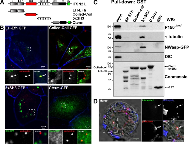
ITSN2 is partially localized at centrosomes through the EH domains and interacts with centrosomal proteins through the SH3 domains. (A) Schematic diagram of the different ITSN2-L domains (called EH-EFh, coiled-coil [CC], 5xSH3, and C terminus) and the GFP and GST constructs generated with them. (B) EH-Efh domains target ITSN2 to the centrosomes. MDCK cell were transfected with GFP-tagged protein constructs of ITSN2 (EH, coiled-coil, 5xSH3, and C terminus) and analyzed by confocal microscopy. Cells were stained to detect pericentrin and actin (blue). Bottom panels show the magnification image of the boxed areas indicated in the top panels. Arrows indicate the localization of EH-GFP to the centrosome (pericentrin) (C) ITSN2 interacts with the centrosomal proteins γ-tubulin and p150Glued. Total cell lysates were incubated with beads preloaded with GST-tagged protein constructs of ITSN2 (EH, coiled-coil, 5xSH3, and C terminus) or with GST. Pulled down fractions were immunoblotted to detect γ-tubulin, p150Glued, N-WASP–GFP (positive control), and DIC or stained with Coomassie blue to detect the total amount of the GST-fused proteins used as bait. Molecular mass is indicated in kilodaltons. WB, Western blot. (D) ITSN2 colocalizes with γ-tubulin and p150Glued in cysts. Cysts expressing vhITSN2 (green) were stained for p150Glued and pericentrin merged with DIC in the left panel. Right panels are the magnification of the boxed area in the left panel. Arrows indicate the colocalization of ITSN2 and p150Glued at the centrosome (pericentrin). Bars, 5 µm.

